Jaipur
+918529833535

'internal temperature control'

Items tagged with 'internal temperature control'

product image
Biochemistry Analyzer

Overview A fully automated POC Auto Dry Biochemistry Analyzer called Mispa Chem Dx, With Just 3 to 4 drops (100ul) of whole blood to test up to 19 General and Special chemistry tests in less than 12 minutes. Mispa Chem Dx works on absorption spectroscopy where a change in absorbance is detected by an absorption spectrum and an algorithm is used to calculate the end concentration of the sample in the reagent dry disc. The reagent disc consists of lyophilized reagent beads, diluent, and calibration details in the QR code. The reagent disc contains an intelligent internal quality control system IQC which continuously monitors the disc function to ensure reaction stability and optimal disc performance, IQC is the engine that drives the Mispa Chem Dx accurately and precisely Low Sample Consumption Easy to Use Maintenance Free Accurate Result Disposable reagent disc Sample volume: 100µl Testing time: 12 minutes / sample Temperature: 37°C ± 0.3°C Resolution: 0.001Abs Absorbance: 0-3.0Abs Sample type: Heparinized whole blood, serum, plasma (heparin) QC&Calibration: IQC (Intelligent Quality Control/Calibration) Work condition: Temperature:10-30°C Humidity: 30%-70% Testing method: End point, kinetic, fixed time, turbidmetry etc Light source: 12V/20W, Halogen tungsten lamp with life span over 2500 hours Power supply: AC 100V-240V, 50-60Hz Display: Android 7.0 inch 800*480, multi-point capacitive touch screen Storage: 500,000 results Printer: Built-in thermal printer Interface: 4 USB ports, 1 LAN port Dimension: 315*375*475mm. 4.2kg(N.W.), 7kg(G.W.)

Send Message
product image
Rapid Test

Buy AccuTest Typhoid IgG/IgM Rapid Card Test (5 Tests) Online from Accurex. The Typhoid IgG/IgM Rapid Test is used for detection & differentiation of anti-Salmonella typhi (S. typhi) IgG and IgM in human whole blood / serum / plasma. This Typhoid test is used as a screening test and as an aid in the diagnosis of infection with S. typhi. Salmonella typhi bacteria causes typhoid Typhoid fever, typhoid symptoms symptoms are likely to develop gradually – appearing one to three weeks after exposure to the disease. There are various typhoid treatment & typhoid vaccine which can be opted after the detetction of Typhoid. Typhoid fever is caused by S. typhi, a Gram-negative bacterium. Worldwide an estimated 17 million cases and 600,000 associated deaths occur annually. Patients who are infected with HIV are at significantly increased risk of clinical infection with S. typhi. Evidence of H. pylori infection also presents an increased risk of acquiring typhoid fever. 1-5% of patients become chronic carrier harboring S. typhi in the gall bladder. The clinical diagnosis of typhoid fever depends on the isolation of S. typhi from blood, bone marrow or a specific anatomic lesion. In the facilities that can not afford to perform this complicated and time-consuming procedure, Filix-Widal test is used to facilitate the diagnosis. However, many limitations lead to difficulties in the interpretation of the Widal test. In contrast, the Typhoid IgG/IgM Rapid Test is a simple and rapid laboratory test. The test simultaneously detects and differentiates the IgG and the IgM antibodies to S. typhi specific antigen thus to aid in the determination of current or previous exposure to the S. typhi. PRINCIPLE The Typhoid IgG/IgM Rapid Test is a lateral flow immunochromatographic assay. The test cassette consists of: 1) a pink-purple colored conjugate pad containing recombinant S. typhi H antigen and O antigen conjugated with colloid gold (Typhoid conjugates) and rabbit IgG-gold conjugates, 2) a nitrocellulose membrane strip containing two test region (Test line M and G) and a control region (control line). The test line ‘M’ is pre-coated with monoclonal anti-human IgM for the detection of IgM anti-S. typhi, test line ‘G’ is pre-coated with monoclonal anti-human IgG for the detection of IgG anti-S. typhi , and the C band is pre-coated with goat anti rabbit IgG. When an adequate volume of test specimen is dispensed into the sample well of the cassette, the test specimen migrates by capillary action across the test cassette. Anti-S. typhi IgM if present in the patient specimen will bind to the Typhoid conjugates. The immunocomplex is then captured on the membrane by the pre-coated anti-human IgM antibody at test region ‘M’, forming a pink-purple line at ‘M’ region, indicating a S. typhi IgM positive test result. Anti-S. typhi IgG if present in the patient specimen will bind to the Typhoid conjugates. The immunocomplex is then captured by the pre-coated reagents on the membrane, forming a pink-purple line at test region ‘G’, indicating a S. typhi IgG positive test result. Absence of any pink-purple line at test region (M and G) suggests a negative result. The test contains an internal control (control line) which should exhibit a pink-purple line of the immunocomplex of goat anti rabbit IgG/rabbit IgG-gold conjugate regardless of the color development on any of the test region. Otherwise, the test result is invalid and the specimen must be retested with another device. PRECAUTION 1. This package insert must be read completely before performing the test. Failure to follow the insert gives inaccurate test results. 2. Do not open the sealed pouch, unless ready to conduct the assay. 3. Do not use expired devices. 4. Bring all reagents to room temperature (15°C-30°C) before use. 5. Do not use the components in any other type of test kit as a substitute for the components in this kit. 6. Do not use hemolized blood specimen for testing. Wear protective clothing and disposable gloves while handling the kit reagents and clinical specimens. Wash hands thoroughly after performing the test. 7. Users of this test should follow the US CDC Universal Precautions for prevention of transmission of HIV, HBV and other blood-borne pathogens. 8. Do not smoke, drink, or eat in areas where specimens or kit reagents are being handled. 9. Dispose of all specimens and materials used to perform the test as biohazardous waste. 10. Handle the Negative and Positive Control in the same manner as patient specimens. 11. The testing results should be read within 15 minutes after a specimen is applied to the sample well or sample pad of the device. Read result after 15 minutes may give erroneous results. 12. Do not perform the test in a room with strong air flow, ie. an electric fan or strong air-conditioning. Other Info STORAGE AND STABILITY Typhoid IgG/IgM test card should be stored at 4-40°C. The card may be stored at room temperature but not exceeding 40°C in the original sealed pouch. The shelf life or expiry of the card is printed on Pouch as well as carton label. The test kit should be kept away from direct sunlight, moisture and heat.

Send Message
product image
Glycohemoglobin Analysis

Buy Xpress Glucometer Plus Kit Online, it’s a smart Glucometer Machine kit. This Digital Glucometer consists of 1 Blood Glucose Meter, 10 Test Strips, 1 Multilet Lancing Device, 10 Sterile Lancets, 1 Carry Pouch, 1 Warranty Card, Test Strip Insert Sheet, 1 Battery (3V) Gluco Plus Meter is intended for use in the home & in clinical settings. There are different types of Glucometers in Point of Care (POC). XPRESSGLUCO + meter measures the strength of the electrical current, calculates your blood glucose level and then displays your result in either milligrams of glucose per deciliter (mg/dL) or millimoles of glucose per liter (mmol/L). The system is intended for use outside the body (in vitro diagnostic use only). It should be used only for self-testing blood glucose (blood sugar) and only with fresh capillary whole blood samples drawn from the fingertips, forearm, upper arm, palm, calf or thigh. The system is intended for use in the home and in clinical settings. The system should not be used for the diagnosis of diabetes or for the testing of newborns. Blood glucose is measured by an electrical current that is produced when a blood samples mixes with the reagent (special chemicals) of the test strip. The electrical current changes with the amount of glucose in the blood sample. The XPRESSGLUCO+ meter measures the strength of the electrical current, calculates your blood glucose level and then displays your result in either milligrams of glucose per deciliter (mg/dL) or millimoles of glucose per liter (mmol/L). Safety Information 1. The user should not take any decision of medical relevance without first consulting his or her medical practitioner. 2. Call your doctor immediately if you experience symptoms that are not consistent with your blood glucose test results. 3. High altitudes above than 3,402 meter (11,161 ft) may affect the test results. 4. Temperatures outside the range of 10°C to 40°C (50°F to 104°F) may affect the test results. Do not test beyond of temperature range. 5. Do not perform servicing and maintenance while the meter is in use. 6. No modification of this equipment is allowed. 7. Do not use this meter near cellular or cordless telephones in a call, walkie-talkies, garage door openers, radio transmitters, or other electrical or electronic equipment that are sources of electromagnetic, radiation, as these may interfere with the proper operation of the meter. Direction for Use 1. Wash you hands and puncture sit: Wash your hands in warm, soapy water. Rinse and dry completely . Warm your fingers to increase blood flow. 2. Insert Test Strip : Remove a new test strip from vial. Be sure to tightly replace vial cap after removing test strips. Insert a test strip with the contact bar end entering into the test strip slot first. Push the test strip as far as it will go without bending it. The meter turns on automatically. 3. Hold the prepared lancing device firmly against the side of your fingertip. Press the release button. (NOTE : If you want to do alternative site testing, please refer to the “About Alternative Site Testing (AST)” section. Please consult your healthcare professional before obtaining blood from site other than your fingertip.) 4. Obtain a Blood Sample : Gently massage your finger or puncture site to obtain the required blood volume. To perform the test, you need only 0.5 μL of blood sample. Do not smear the blood sample. To obtain best accurate result, wipe off the first drop of blood and gently squeeze another drop of blood. 5. Apply Blood Sample : When the meter shows the “ ” symbol, apply blood to the opening of the absorbent channel of the test strip where it meets the narrow channel. Blood will be drawn into the test strip. If the test strip confirmation window is full, you will hear a beep. 6. Read Your Result : After the meter counts down from 5 to 1, your blood glucose test result appears along with the unit of measure, date and time. Test result is lower than the target range Test result is within the target range. Test result is higher than the target range *The default target range is 70 mg/dl to 180 mg/dl. 7. Turn Off the Meter : This blood glucose result is automatically stored in the meter memory. Turn the meter off by removing the test strip. Discard the used test strip carefully to avoid contamination. 8. After use, twist off the Lancing Device Cap. Push the exposed tip of the lancet into its Protective Cap. 9. Slide the Lancet Ejector forward and disposing the used lancet in an approved container. Discard the used lancet according to your country’s safety regulations. Replace the Lancing Device Cap. Other Info •Use only XPRESSGLUCO+ Control Solution with your XPRESSGLUCO+ meter. •Check the expiration date on the bottle. Do not use if expired. •Use within a period of 90 days from the date that you first open it. Record the discard date on the control solution bottle when you first open it to serve as a reminder to discard after 90 days. •The control solution ranges are printed on the label of the XPRESSGLUCO+ Blood Glucose Test Strip vial. They are not recommended target ranges for your blood glucose. •For in vitro diagnostic use. •Do not add any liquid to the XPRESSGLUCO+ Control Solution. •Do not take internally or inject.

Send Message
product image
Hematology

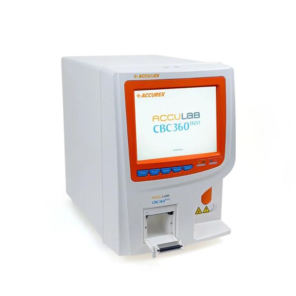
Buy the Best Automated Hematology Analyzers – Acculab CBC 360 Neo. This Hematology Analyser Machine is Truly Automated for self-checking, background test, calibration, sampling, dilution, mixing, printing, dormancy, alarms, cleaning and maintenance. CBC 360 Neo is 3 part hematology analyzer from Accurex & is Automated for Walk-away Operations. Dual Chamber: Measurement of RBC / platelets & WBC / HGB in different chambers ensure accurate counting in a short time. The micro-aperture sizes of both the chambers are well designed to avoid error due to cell misclassification Automation for Walk-away Operation: Auto Self-check : Automatic aspiration of reagents & rinsing of tubings on startup Auto Blank : Background test is run automatically on startup Auto Print : Automatic printing of report with / without histogram Auto Sleep : Dormancy status is entered after a set period of non-operation Auto Alarms : Audio / visual alarms in case of system errors & abnormal results Auto Clean : Automatic rinsing options available to maintain the system Dual Chamber: Measurement of RBC / platelets & WBC / HGB in different chambers ensure accurate counting in a short time. The micro-aperture sizes of both the chambers are well designed to avoid error due to cell misclassification Specifications: Measurement principle: Electrical resistance for counting RBC, WBC & Platelets Colorimetric estimation of HGB at 540 nm Parameters: WBC, LY, MO, GR, LY%, MO%, GR%, RBC, HGB, HCT, MCV, MCH, MCHC, RDW-CV, RDW-SD, PLT, MPV, PDW, PCT, P-LCR Throughput: 60 samples/hour Micro-aperture: WBC : 100 um RBC / PLT : 68 um Sample type: Whole blood or pre-diluted sample Sample volume: Whole blood (venous blood) : 10 ul Chamber: Pre-diluent (capillary blood) : 20 ul, Pre-diluent (capillary blood) : 20 ul Reagents: Dual chamber : RBC / Platelets & WBC / HGB Quality control: 4 QC options : L-J, X-B, X-R & X QC, Graphs plotted with 31 data points Data memory: Upto 100,000 (with histogram) Display: 10.4 inch LCD with 640 x 480 resolution Printer: Built-in thermal printer Interface: Support RS-232C, standard network port and USB; keyboard and mouse Operating environment: Temperature : 15˚C – 35˚C, Humidity : 85% RH Electrical specification: Voltage : AC 110V/230V 23V Frequency : 50/60 Hz, Power : 180W Fuse specification : 250V/3A Dimensions / Weight: 670 x 515 x 640 mm / 27.5 kgs Specification Acculab CBC 360 Neo – Fully Automated Hematology Analyzer Features: 3-part differentiation of WBC Automated for — self-checking, background test, calibration, sampling, dilution, mixing, printing, dormancy, alarms, cleaning and maintenance High processing speed : 60 samples/hour Multi-parameter estimation : 20 parameters categorized according to corpuscle type for easy reporting Direct command keys for Mode, Prime, Flush, Drain & Record / Print for faster operation Run / Standby indicator lights denote the analyzer status i.e. test is being run or analyser is ready to test Whole blood/ Pre-diluent indicator lights denote the sample type in use i.e. whole blood/ pre-diluent Dual sample mode with low sample volume: Whole blood (venous) mode : 10 ul; pre-diluent (capillary) mode : 20 ul Audio alarm indication for abnormal findings or system errors 4 QC methods : L-J, X-B, X-R & X QC Comprehensive QC graphs with 31 data points for each parameter Internal & external probe cleaning facility Dormancy mode available to reduce power consumption Memory : 100,000 (with histogram) Safety Information • Large 10.4 inch single screen color display of all 21 parameters and 3 histograms • Sequential arrangement of WBC, RBC & PLT parameters for convenient reporing •Easy to understand & icon-based arrangement of menu for fast and convenient testing Direction for Use • WBC Histogram: R1, R2, R3, R4 & RM indications on histogram show specific abnormalities in the histogram & the probable causes • Platelet Histogram: PM indication is given when the boundary between PLT & RBC is ill-defined thus avoiding misclassification Other Info High voltage cautery function is useful in disintegrating obstinate protein or serum clog in the tubing

Send Message
product image
Immunology Analyzer

Product Description: The LisaScan EM Microplate Reader is an economical, compact and versatile 8 channel ELISA reader which can be adapted to various ELISA applications. It is India’s largest selling automated microplate ELISA reader Product Features: Wavelength Range: 400 to 750nm with a dynamic measurement range of 0.0 to 4.0 O.D 100 user-programmable test storage Inbuilt thermal printer for reports & graphs Auto self-check on start-up & external printer connectivity Compatible to various plate geometrics-96 well microplates ( Flat, U & V bottom) Wavelength selection: Monochromatic, bi-chromatic & multi-chromatic* Reading time 8 sec. for single wavelength using 8-channel optics User-friendly inbuilt operation software with 7000 sample test results storage User interface: High Resolution LCD display with 20 Keys Rugged waterproof & membrane panel Computer linked ELISA data management software - Eli-LIMS* CE marked Inbuilt shaking facility,Three Linear Speeds - Low, Medium and Fast Reports can be printed using internal thermal or external printer No need of manual calculations and documentation Documentation ease – Reports can even be stored using optional Eli-LIMS software Computer can be connected Extensive ELISA data management Product Applications: ELISA System type: Open System Plate types: 96 well plates Wells Type: U, V and Flat bottom wells Operating Modes: Absorbance Quantitative Qualitative Semi-Quantitative Kinetic Mode Measurement Modes: Continuous / Step Shaking Modes: Three Linear Speeds - Low, Medium and Fast. Time duration can be set Wavelength Selection: Monochromatic Bi-Chromatic Multi-Chromatic* (Maximum 4) Calculation Modes: Non-linear Standard Cut-off Index Formula Column Subtraction Linear Regression Point to Point Cubic Spline Linear – Log Blank Options: Blank, Control, Assay Validation equations Against Air, Well Plate, Column, Column-mean Row, Row mean Number of programmable standards / calibrators: Up to 10 standards /test Storage/ display/ printing of multi standard curves and cut-off equations for all parameters Test Programs: 100 totally “open” Test Programs Photometer: Measuring system: 8-Channel optical system Lamp Source: Tungsten Halogen with Lamp saver function Wavelength Range: 400 to 750 nm Standard Filters: 405 nm, 450 nm, 492 nm, 630 nm (Optional two filters 578 nm, 700 nm), 10 nm Band Pass Dynamic Range: 0.0 ~4.0 OD Photometric accuracy: < 1% at 2.5 OD at 450 nm Photometric resolution: 0.001 OD Linearity: <1% at 2.5 OD at 450 nm Measurement Time: SPEED : 8 Seconds single wavelength NORMAL : 17 Seconds single wavelength Display: High resolution Graphics LCD 320x240 pixels, LED Backlight, View area: 120x92 mm Keyboard: Rugged waterproof & membrane panel PC Communication: USB Based Protocol compatible with Eli-LIMS and TNW Built-in Thermal Printer: Built in High resolution, 384 dots per line, thermal Type with full graphics facility printer and option for connecting External Parallel Printer (or USB Printer using External Adaptor). Computer interfacing Software: Windows Based Software Eli-LIMS with data managemenOptional Windows Based Software with data management capabilities to store patient results, store/display calibration curve /QC curves. Reports on internal and external printers: Report generation for last 7000 Sample’s is viewed/printed/stored. Lab name in Print-out Lab name in Display Collated reports Print-out of Normal Range along with the result (on external printer only). Graphs can be printed on Thermal Printer Sorted by: Date I.D. Both by Date & I.D. Test Plate ID Online Help: At each step on-line help in each menu. Beeps and online instructions on erroneous entry. Special HELP key on keypad. Operating Temperature: 20˚ C to 40˚ C Storage Temperature: -10˚ C to 50˚ C Humidity: Max. 80% RH, non- condensing Power Supply: 16 V - 24.5 V DC using External Auto ranging SMPS adapter 115/230VAC ±10%, 50/60Hz., Max. 90 Watt. (CE & UL certified) Dimension: 502 mm X 369 mm X 216 mm Weight: 7 Kg

Send Message
product image
Hematology

VES MATIC 5 is the next-generation automated module manufactured by Diesse Diagnostica (Italy), a highly advanced, Intelligent, Intuitive & Interactive Graphic user Interface for estimating ESR using EDTA samples. Product Features: Automated instrument for the determination of ESR directly from EDTA vacutainers Faster TAT by loading 216 sample tubes at a time Pediatric samples can be processed in microtainers Reliable results with Temperature correction facility Accurate results by eliminating manual HCT correction in Anaemic samples Real-Time monitoring of samples Detection of Lipemic, Hemolyzed, Coagulated samples Standardization in mixing with Inbuilt Mixer Analyser P. Code Product Name Pack size Vesmatic 5 165941 Next 5K Transponder 1x 5000 Vesmatic 5 165942 Next 10 K Transponder 1x 10000 Display - 19" Full HD (1920x1080) LCD with PCAP Touch Screen Peripheral Control - based on Everex distributed control system with peripheral motor control boards Operating system - Linux Analysis module - 89 Positions for Sample Tubes Sample Loading - up to 18 racks simultaneously Throughput - 190 samples per hour. First result after 28 minutes Walk away mode - Supported Continuous loading - Supported Sample mixing - with multiple 180° rotations of the tubes Collection of analysed samples - reinserted into the original loading rack LIS Connection - Multiple LIS connection protocols are available: ASTM / Proprietary Remote diagnostics - available via Ethernet connection. Printer -Alphanumeric with 58mm-wide thermal paper, 36 characters per line, speed 20 mm/sec. Imaging System - 2 high resolution cameras for sample identification and analysis; 1 standard camera for internal inspection Image processor - Dedicated Quad Core CPU with integrated GPU for real-time HD image processing of samples Temperature range in operation - from +15 to + 35°C; Storage from + 5°C to + 45°C Humidity range - from 20% to 80% without condensation Central unit - Quad core ARM Cortex A-53 CPU ARM Cortex A-53, 16 GB eMMC, 4 GB RAM Interfaces - 2 x RS232C, 2 Host USB, 1 Client USB, Ethernet Protection category - CLASS I Safety standard - EN 61010-1, EN 61010-2-081, EN 61010-2-101 EMC - EN61326-1 Installation category - II Mains voltage - 230VAC@50Hz; US/Canada: 110-120Vac@60Hz Electrical energy consumption - 420VA Fuse block - 2 x 5.0 AT (delayed) (5 x 20 mm) UL Dimensions (mm) - 850 (W) x 750 (H) x 830 (D) Weight - 80 kg

Send Message
product image
immunoassayReagents

H-FABP Rapid Quantitative Test Finecare™ H-FABP Rapid Quantitative Test is a fluorescence immunoassay for quantitative measurement of Heart-type Fatty Acid Binding Protein (H-FABP) in human whole blood, serum or plasma. It is used for diagnosis of myocardial infarction.With original qualify control. STORAGE AND STABILITY 1. Store the test kit at 4℃~30℃ up to the expiration date printed on package. 2. If removed from refrigerator, allow the test kit for 30 minutes to return to room temperature before testing. 3. Do not remove the Test Cartridge from the pouch until use. The Test Cartridge should be used within 1 hour once opened.

Send Message
product image
Clinical Chemistry

With cutting-edge LED technology, the HyLED 7 Series offers excellent luminous efficiency, heat-free light source, and incredible long service life, giving you total support in OR. Usage/Application Operation Theater Positioning Ceiling Mounted Model Name/Number HyLED 7 Series Brand Mindray Light Source Type LED Central illuminance at 1m distance 160,000 lux Light field diameter(at 1m distance) 180~300 mm Depth of illumination(L1+L2) 1,200 mm Correlated color temperature 4,350 K Color rendering index(Ra) 96 Color rendering index(R9) 97 Shadow dilution with tube 100% Shadow dilution with one mask 75% Shadow dilution with tube and one mask 70% Shadow dilution with two masks 55% Shadow dilution with tube and two masks 50% Ambient illumination 8,000 lux Bulb power consumption 85 W Power supply 100~240 VAC, 50/60 Hz Number of LED bulbs 32 Light head dimension 600 mm Integrated Camera Optional Carrier-arm Camera Optional HyLED 7 Series Contact Us With cutting-edge LED technology, the HyLED 7 Series offers excellent luminous efficiency, heat-free light source, and incredible long service life, giving you total support in OR. Efficient & Economic • Incredible long service time up to 60,000 hours • Central Illumination: 160,000 lux/130,000 lux • Adjustable light field diameter • Max. depth of Illumination : 1,200 mm • Low power consumption Original & Unique • Perfectly integrated into laminar flow (Certified per DIN-1946 Part 4) • Touch screen panel for easier control • 330° rotatable integrated HD/FHD camera • Ultra-thin design & excellent maneuverability • Ergonomic and compact design less than 12kg Ergonomic and Compact Design • The ultra-thin concentrically circular design for better hygiene. • DIN-1946 Part 4 certificated. (12-2008) Flexible Design • Combined with an Ondal spring arm maximizes mobility and ensures stability. • Ultra-thin light head with only less than 12 Kg. • Smooth, metal back cover of the light head. High Performance • With a minimum lifetime of 60,000 hours, each latest generation LED illuminates far longer than traditional light sources. • Easier and less costly maintenance: each LED bulb with individual circuit can be replaced respectively, which will minimize the maintenance cost. Easy and Intuitive Control • Traditional control panel • Touch screen panel(optional) Shadow-free The HyLED 7 Series surgical light inherits the advanced technology from the HyLED 9 series and shows excellent shadow management function. The professional shadow dilution experiment simulated extreme situations by using tubes and masks proved that Mindray HyLED 7 Series surgical light has sufficient illumination even in extreme situations. Camera System for Communication The camera system provides better communication and documenting solutions. The camera system integrated with the surgical lights also provides a platform for medical research purpose, teaching communication and video documenting. Digital video recorder Supporting external USB storage device Supporting HDMI/YPbPr/HD-SDI/DVI-D/VGA input signal Default with camera control function Supporting video recording from other medical devices source

Send Message
product image
Medical Equipment

Since the company was founded, Mindray has been continuously exploring new ways to improve diagnostic confidence. Powered by the most revolutionary ZONE Sonography® Technology, Resona 7’s new ZST+ platform brings ultrasound image quality to a higher level by zone acquisition and channel data processing. As well as the premium level image quality, Resona 7 also enhances clinical research capabilities with the revolutionary V Flow for vascular hemodynamic evaluation, and the most intelligent plane acquisition from 3D datasets for fetal CNS diagnosis. Combining the most intuitive gesture-based multi-touch operation and all the essential clinical features, Resona 7 is truly leading new waves in ultrasound innovation. With core platform advantages of ZST+ The channel data based ZST+ is an extraordinary innovation, representing an ultrasound evolution. Transforming ultrasound metrics from conventional beamforming to channel data based processing, ZST+ is able to deliver multiple imaging advances: Advanced Acoustic Acquisition, Dynamic Pixel Focusing, Sound Speed Compensation, Enhanced Channel Data Processing and Total Recall Imaging. Advanced features V Flow V Flow (Vector Flow) is a novel approach for vascular hemodynamic analysis. V Flow uses color coded vector arrows to indicate the velocity’s magnitude and direction of blood cells. With ultra-high frame rates, it provides extremely accurate and angle-independent visualization of complex vascular hemodynamic profiles. With comprehensive data information, V Flow is the most valuable tool for vascular clinical research. resona-7-fig3-1-pc UWN+ Contrast Imaging UWN+ (Ultra-Wideband Non-linear Plus) CEUS enables the Resona 7 to detect and utilize both the 2nd harmonic and non-linear fundamental signals, generating significantly enhanced images, resulting in greater sensitivity of minor signals and longer agent duration with lower MI. Forwarding smart to clinical intelligence The Resona 7 elevates clinical intelligence with a complete solution that enables clinicians to manage both routine and advanced studies more efficiently,consistently, and accurately, from acquisition to calculation. As an example, Smart Planes CNS shows exceptional intelligence in accurate diagnosis and analysis of the fetal central nervous system (CNS) Smart Planes CNS Mindray’s exclusive pioneering technology positions the Resona 7 as the industry’s first ultrasound system to allow fully automatic and accurate detection of the most significant planes and frequently used measurements of fetal CNS, leading to intelligent diagnosis, improved throughput, and reduced user dependence. Smart Planes CNS provides a user-friendly tool that greatly improves scanning efficiency through increased accuracy coupled with automated operation. With a simple button click on a 3D fetal brain volume image, the standard CNS scanning planes (MSP, TCP, TTP and TVP) and a range of related anatomical measurements (BPD, HC, OFD, TCD, CM and LVW) are obtained immediately. Smart Face Acquiring an optimal view of the fetal face in 3D ultrasound is cumbersome and time-consuming. In some cases, it is impossible to get rid of the occlusions such as cord, placenta, uterus and extremities. The new Resona 7 with Zone Intelligence provides a fast and intelligent optimization for fetal face with simply one-touch. It can immediately remove occlusions in the volume data, eliminate unwanted noise information, and generate an optimal view of the fetal face with minimal effort. Ergonomics The Resona 7 is designed around you. Gesture-based operation opens up a new trend in cart-based ultrasound with an agile, smart, and intuitive user experience beyond your expectations. Gel warmer’s three level temperature and swiveling angle adjustment of the control panel delivers great patient comfort and user convenience.

Send Message

Still searching for
internal temperature control?