Jaipur
+918529833535

'epithelial cells'

Items tagged with 'epithelial cells'

product image
Hematology

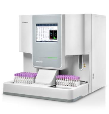
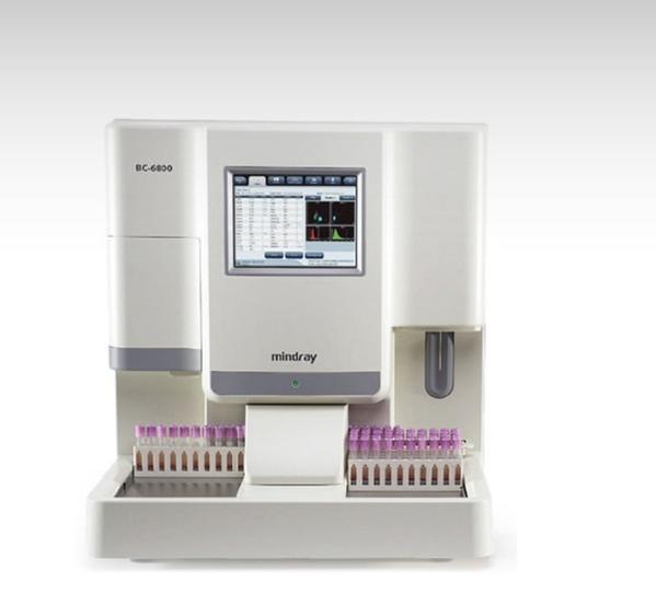
BC-6800 Today, laboratories not only need more reliable routine CBC plus 5-part diff WBC testing by way of high processing speed, but are also looking for options that extend the analyzer's output in form of Reticulocytes, Nucleated RBCs, Fluorescent Platelet counts etc. for wider clinical application and research. On the other hand, they are restricted by limited budgets. BC-6800 can help to meet all these needs just perfectly and even exceed the expectations. Additional parameters for further clinical applications IMG(#,%) parameters provide information about immature granulocyte, including promyelocytes, myelocytes and metamyelocytes. HFC*(#,%) parameters represent high fluorescent cell population, such as blasts and atypical lymphocytes. RET(#,%) extend valuable help in the differential diagnosis and/or therapeutic monitoring of anemias. IRF is parameter concerning immature reticulocytes, which can assist in early diagnosis of anemia and monitoring the bone marrow response to therapy. InR*(#,‰) are parameters regarding infected red blood cell. The red blood cells may infected by plasmodium, which can cause malaria. * means research parameters SF Cube Technology SF Cube is a pathbreaking technology for reliable blood cell analysis, including WBC differential, Reticulocytes and NRBC with efficient flagging. After reaction with proprietory reagents, the targeted blood cells undergo 3D analysis using information from scatter of laser light at two angles and fluorescence signals. The 3D scattergram builds the power to better identify and differentiate blood cell populations, especially to reveal abnormal cell population undetected by other techniques. Minimizing interference to ensure more accurate results NRBCs are counted in a dedicated channel by SF cube method, which automatically corrects total WBC count and 5-part Diff results when NRBCs are detected. The Focusing Flow-DC method minimizes the interference traditionally encountered in DC technology to produce near gaussian histograms. PLT-O result avoids the interference from microcytic and fragmented RBCs, large platelets and/or platelet clumps by fluorescent stain, and enhances the results accuracy and sensitivity. High automation to minimize the workload Throughput for 125 tests/hour, autoloader for 100 sample tubes capacity Bi-directional LIS with HL7 or ASTM communication protocol Customizable re-exam rules allow users to define their re-exam criterion according to lab’s practice

Send Message
product image
Hematology

The advanced BC-6800Plus not only provides more reliable routine CBC plus 5-part DIFF results, but also has a higher throughput of up to 200 samples per hour. Based on the new SF Cube technology platform, the BC-6800Plus could provide more reliable blood cell results and abnormal cells flagging results with anticipated warning messages, exceeding clinicians’ expectations and setting a new standard for blood analysis. SF Cube Technology With the newly designed opticals and reagent systems, the SF Cube technology can help doctors to better differentiate the clusters of cells, which is the key to revealing more abnormal cells. NRBC result in every CBC In WNB scattergram, BC-6800Plus provides NRBC, Basophils and WBC-N* results. It means that the actual number of NRBCs can be measured in routine CBC, if they are present in the sample. Basophils are counted in this counting channel with NRBC results. Basophil and NRBC results are generated on BC-6800Plus without extra reagent or cost. NLR parameter in each CBC+DIFF Neutrophil-to-lymphocyte ratio (NLR) is calculated by dividing neutrophil count by lymphocyte count, usually from peripheral blood sample. In laboratory medicine, NLR is used as a marker of subclinical inflammation. According to clinical studies, NLR can be used as a prognostic factor of severe illness in COVID-19 patients. More accurate RET and PLT-O result By utilizing the new fluorescent dye, the reticulocytes and platelets are more specific stained with stronger fluorescent signal, that brings more reliable reticulocyte and platelet results. Automatic Rerun & Reflex Should the sample results trigger the criteria, the sample rack can automatically step back for a “rerun” or “reflex” check. 200 test/hour BC-6800Plus can load up to 100 samples at a time and offers a throughput of up to 200 tests per hour.

Send Message
product image
Hematology

n large laboratories, rows of instruments are seen working tirelessly like miniature factories – only they produce not commodities but medical test results. Mindray CAL 8000 new-generation cellular automation line is one of them, but unlike a typical workhorse, it is intelligent. It is a highly automated workstation for smarter, faster, and more accurate cellular analysis. It can perform competently in the face of these challenges. The workstation can hold up to four units of BC-6800 automated hematology analyzer, which utilizes the advanced SF Cube technology and 3-D scatter grams to help doctors better differentiate blood cell populations and reveal abnormal cells undetectable by conventional techniques. With 125 CBC tests per hour per unit, BC-6800 brings the process rate of the workstation to 20–30 percent higher than other similar models, effectively alleviating laboratory's stress during the peak processing hours. On CAL 8000, the hematology analyzer, slide maker and stainer, CRP analyzer, and HbA1c analyzer are all integrated into one seamless production line through automated track modules and trolley. Simply by loading a single tube of blood sample onto the automation system, one can get all the test results. This innovative walk-away automation functionality has made possible the CBC+CRP+HbA1c testing all in one streamlined workstation for the first time in the industry. It offers a smart solution, or rather, a smart expert – the labX pert 2.0. It is a software that can automatically sort out the suspected samples for manual validation. The pathologist then decides if rerun or microscopic review is warranted based on their clinical experience. It avoids wasting slides and reduces extra work. Moreover, the onboard automated re-exam functionality also helps enhance work efficiency. The CMU software The touch screen is the “brain” of CAL 8000. It optimizes the distribution of workload between each analyzer unit and decides which sample requires a blood smear. It displays the status of each analyzer or SC-120 installed on CAL 8000 as well as the volume of balance reagent. This intuitive software can tell the users all necessary information of CAL 8000 at any given time. Consequently, the users spend less time operating CAL 8000 and can arrange their working schedule better. Multi “R” tests The CAL 8000 can automatically distribute the samples with “Repeat”, “Rerun” and “Reflex” criteria which are pre-defined by users.The re-exam criteria on CAL 8000 can be defined by many conditions, such as time, patient’s age, gender, department, etc. This helps minimize the number of blood smears. Special tube racks The special tube racks utilize different barcodes to differentiate specific testing purposes, such as “QC”, “Slide making & staining only” and “RET test only”. The special tube racks can carry the tubes to perform these specific functions in the simplest way.

Send Message
product image
Medical Equipment

Since the company was founded, Mindray has been continuously exploring new ways to improve diagnostic confidence. Powered by the most revolutionary ZONE Sonography® Technology, Resona 7’s new ZST+ platform brings ultrasound image quality to a higher level by zone acquisition and channel data processing. As well as the premium level image quality, Resona 7 also enhances clinical research capabilities with the revolutionary V Flow for vascular hemodynamic evaluation, and the most intelligent plane acquisition from 3D datasets for fetal CNS diagnosis. Combining the most intuitive gesture-based multi-touch operation and all the essential clinical features, Resona 7 is truly leading new waves in ultrasound innovation. With core platform advantages of ZST+ The channel data based ZST+ is an extraordinary innovation, representing an ultrasound evolution. Transforming ultrasound metrics from conventional beamforming to channel data based processing, ZST+ is able to deliver multiple imaging advances: Advanced Acoustic Acquisition, Dynamic Pixel Focusing, Sound Speed Compensation, Enhanced Channel Data Processing and Total Recall Imaging. Advanced features V Flow V Flow (Vector Flow) is a novel approach for vascular hemodynamic analysis. V Flow uses color coded vector arrows to indicate the velocity’s magnitude and direction of blood cells. With ultra-high frame rates, it provides extremely accurate and angle-independent visualization of complex vascular hemodynamic profiles. With comprehensive data information, V Flow is the most valuable tool for vascular clinical research. resona-7-fig3-1-pc UWN+ Contrast Imaging UWN+ (Ultra-Wideband Non-linear Plus) CEUS enables the Resona 7 to detect and utilize both the 2nd harmonic and non-linear fundamental signals, generating significantly enhanced images, resulting in greater sensitivity of minor signals and longer agent duration with lower MI. Forwarding smart to clinical intelligence The Resona 7 elevates clinical intelligence with a complete solution that enables clinicians to manage both routine and advanced studies more efficiently,consistently, and accurately, from acquisition to calculation. As an example, Smart Planes CNS shows exceptional intelligence in accurate diagnosis and analysis of the fetal central nervous system (CNS) Smart Planes CNS Mindray’s exclusive pioneering technology positions the Resona 7 as the industry’s first ultrasound system to allow fully automatic and accurate detection of the most significant planes and frequently used measurements of fetal CNS, leading to intelligent diagnosis, improved throughput, and reduced user dependence. Smart Planes CNS provides a user-friendly tool that greatly improves scanning efficiency through increased accuracy coupled with automated operation. With a simple button click on a 3D fetal brain volume image, the standard CNS scanning planes (MSP, TCP, TTP and TVP) and a range of related anatomical measurements (BPD, HC, OFD, TCD, CM and LVW) are obtained immediately. Smart Face Acquiring an optimal view of the fetal face in 3D ultrasound is cumbersome and time-consuming. In some cases, it is impossible to get rid of the occlusions such as cord, placenta, uterus and extremities. The new Resona 7 with Zone Intelligence provides a fast and intelligent optimization for fetal face with simply one-touch. It can immediately remove occlusions in the volume data, eliminate unwanted noise information, and generate an optimal view of the fetal face with minimal effort. Ergonomics The Resona 7 is designed around you. Gesture-based operation opens up a new trend in cart-based ultrasound with an agile, smart, and intuitive user experience beyond your expectations. Gel warmer’s three level temperature and swiveling angle adjustment of the control panel delivers great patient comfort and user convenience.

Send Message
product image
Rapid Test

Buy AccuTest HIV – 1/2 Ab Rapid Card Test (50 Tests) Online from Accurex. This Rapid HIV test is a visual, solid phase immunochromatographic assay for qualitative in-vitro diagnostic detection of antibodies against HIV-1 and HIV-2 in human serum/plasma. This HIV test may be used as an aid in diagnosis of HIV infection. In the HIV blood test – A reactive result should be confirmed by supplemental testing as part of a validated HIV testing algorithm. INTENDED USE: AccuTest HIV – 1/2 Ab test is a visual, solid phase immunochromatographic assay for qualitative in-vitro diagnostic detection of antibodies against HIV-1 and HIV-2 in human serum/plasma. The product may be used as an aid in diagnosis of HIV infection. A reactive result should be confirmed by supplemental testing as part of a validated HIV testing algorithm. INTRODUCTION: The Human Immunodeficiency Virus (HIV) is a retrovirus that infects cells of the immune system, destroying or impairing their function. The most advanced stage of HIV infection is Acquired Immunodeficiency Syndrome (AIDS). The general method of detecting infection with HIV is to observe the presence of antibodies to the virus by an EIA method followed by confirmation with any confirmation method. AccuTest HIV – 1/2 Ab is a simple, visual qualitative test that detects antibodies in human serum/plasma. The test is based on immunochromatography and can give a result within 15 minutes. PRINCIPLE: AccuTest HIV – 1/2 Ab employs chromatographic lateral flow device in a cassette format. Colloidal gold conjugated recombinant antigens (Au-Ag) corresponding to HIV-1 (gp120 + gp41) and HIV-2 (gp-36) are dry-immobilized at the end of nitrocellulose membrane strip. HIV-1 & HIV-2 antigens are bound at the Test Zone (1 & 2) respectively. Goat Anti-Mouse IgG antibodies are bound at the Control Zone(C). When the sample is added, it migrates by capillary diffusion rehydrating the gold conjugate. If there are HIV-1 or HIV-2 antibodies in sample, they will bind with the gold conjugated antigens forming particles. These particles will continue to migrate along the strip until the Test Zone (1 or 2) where they are captured by the HIV-1 or HIV-2 antigens generating a visible purple line. If the specimen contains antibodies to HIV-1, the specimen will bind to the 1 test line and produce line. If specimen contains antibodies to HIV2, the specimen will bind to the 2 test line. If there are no HIV-1 or HIV-2 antibodies in sample, no purple line is formed in the Test Zone. The gold conjugate will continue to migrate alone until it is captured in the Control Zone (C) by the Goat Anti Mouse IgG antibodies aggregating in a purple line, which indicates that the migration of liquid has been successful. The control line must be present for the test to be valid. PRECAUTIONS: 1. For best results, adhere to instructions provided. 2. All specimens should be handled as potentially infectious. 3. The test device is sensitive to humidity as well as heat. 4. Do not use beyond expiration date. 5. Do not use test kit if pouch is damaged or seal is broken. 6. Use test device immediately after removing from the pouch. 7. The components (test device and assay diluents) in this kit have been quality control tested as a standard batch unit. Do not mix components from different lot numbers. 8. The AccuTest HIV – 1/2 Ab Test devices should be stored at 4 to 30 °C. Do not expose the kit to temperature over 30 °C. 9. Specimen with extremely high concentrations of red blood cells, fibrin should be recentrifuged before use. 10. AccuTest HIV – 1/2 Ab Test Device (Serum/Plasma) is for in vitro diagnostic use only. 11. AccuTest HIV – 1/2 Ab Test Device (Serum/Plasma) will only indicate the presence of antibodies to HIV-1/2 in the specimen and should not be used as the sole criteria for the diagnosis of HIV-1, HIV-2, and/or HIV-1 subtype O infection. 12. For confirmation of reactive test results, specimens should undergo further testing using different assays, such as rapid diagnostic tests, EIA and/or Western blotting in accordance with a validated HIV testing algorithm. 13. As with all diagnostic tests, all results must be interpreted together with other clinical information available to the physician. 14. Results should not be used to determine the serotype of HIV infections TEST PROCEDURE: 1. Allow the test device, buffer and specimen to reach room temperature (15-30 °C) prior to testing. 2. Remove the test device from the foil pouch and use the test device as soon as possible (within one hour). 3. Place the device on a flat, dry surface. Label with specimen ID. 4. Holding the sample dropper vertically above the test disk, slowly transfer 1 drop (10µl) of sample in to the sample well, then add 2 drops of assay diluent (about 75 µl) and start the timer. 5. As the test begins to work, you will see purple color moving across the result Window in the center of the Test Disk. 6. Wait for the colored lines to appear. Interpret test results at 15 to 20 minutes. Do not interpret test result after 20 minutes. STORAGE AND STABILITY Store as packaged in the sealed pouch at 4-30 °C (storage in refrigerator is permitted). Do not store in the freezer. Protect the test kit from humidly. The test device is stable until the expiration date printed on the test kit and/or sealed test device pouch. Do not use beyond the expiration date. The test device must remain in the sealed pouch until use.

Send Message

Still searching for
epithelial cells?