Jaipur
+918529833535

'accurate diagnosis'

Items tagged with 'accurate diagnosis'

product image
Rapid Test

Intended Use Hepacard is visual, rapid, sensitive and accurate one step immunoassay for the qualitative detection of Hepatitis B surface antigen (HBsAg) in Human serum or plasma.The assay is intended to be used as an aid in the recognition and diagnosis of acute infections and chronic infectious carriers of the Hepatitis B Virus(HBV). Salient Features Detects all the 11 subtype of HBsAg. Antigen Sensitivity- 0.5 ng/ml. No sample preparation required. See through Device for easy result interpretation. Shelf life-30 months at 2-30°C. WHO Evaluated with 100% Sensitivity.

Send Message
product image
Rapid Test

Intended Use Diagnos Celiac Card is a visual, rapid, sensitive and accurate one step immunoassay for the qualitative detection of anti tTG IgA, IgG & IgM antibodies in Human Serum or Plasma. The assay is intended to be used as an aid in the recognition and diagnosis of Celiac disease. Salient Features Rapid, Visual and Qualitative Antibody Test. Excellent Sensitivity: 99.26% & Specificity: 99.50%. Results within 20 minutes. Long Shelf life 24 months at 2-8°C. Bio-hazard free, fully covered, see-through Device. No instrument required.

Send Message
product image
Clinical Chemistry

Designed to fulfill the clinical requirements of minimally invasive surgeries, Mindray's HD3 endoscope camera system comes with the advanced 3-chip CMOS sensor, premium optical lens, real-time optimization software and a FHD screen. Through delivering exceptional image quality and user experiences, the HD3 endoscope camera system enables surgeons to focus on their patients, helping facilitate more accurate diagnosis and treatments. HD3 Camera System 3-chip CMOS sensor FHD picture taking and video recording with USB port Advanced parfocal technology 2X optical zoom Focal length: 14mm-30mm Laparoscopes · High-resolution images and outstanding color rendition · Scratch-resistant sapphire lens · Fog reduction function · Autoclavable lens · Compatible with third-party optical fiber cables HS50 Insufflator · Maximum flow rate: 50L/min · Large 7-inch touchscreen · Gas heating function · Smoke evacuation · Automatic flow compensation · Auto-switchover between veress needle mode and trocar mode · Up to 5 insufflation modes, satisfy all insufflation needs HB200L LED Light Source · Equivalent to 300W Xenon light source · Autoclavable light cable · Auto-detection of light guide cable · Light intensity indicator · LED life expectancy: 40,000 hours PV27 FHD Monitor · 1920*1080 resolution · Picture-in-picture mode · Adjustable brightness · Flexible input: DVI,SDI,S-video,VGA

Send Message
product image
Hematology

BC-6800 Today, laboratories not only need more reliable routine CBC plus 5-part diff WBC testing by way of high processing speed, but are also looking for options that extend the analyzer's output in form of Reticulocytes, Nucleated RBCs, Fluorescent Platelet counts etc. for wider clinical application and research. On the other hand, they are restricted by limited budgets. BC-6800 can help to meet all these needs just perfectly and even exceed the expectations. Additional parameters for further clinical applications IMG(#,%) parameters provide information about immature granulocyte, including promyelocytes, myelocytes and metamyelocytes. HFC*(#,%) parameters represent high fluorescent cell population, such as blasts and atypical lymphocytes. RET(#,%) extend valuable help in the differential diagnosis and/or therapeutic monitoring of anemias. IRF is parameter concerning immature reticulocytes, which can assist in early diagnosis of anemia and monitoring the bone marrow response to therapy. InR*(#,‰) are parameters regarding infected red blood cell. The red blood cells may infected by plasmodium, which can cause malaria. * means research parameters SF Cube Technology SF Cube is a pathbreaking technology for reliable blood cell analysis, including WBC differential, Reticulocytes and NRBC with efficient flagging. After reaction with proprietory reagents, the targeted blood cells undergo 3D analysis using information from scatter of laser light at two angles and fluorescence signals. The 3D scattergram builds the power to better identify and differentiate blood cell populations, especially to reveal abnormal cell population undetected by other techniques. Minimizing interference to ensure more accurate results NRBCs are counted in a dedicated channel by SF cube method, which automatically corrects total WBC count and 5-part Diff results when NRBCs are detected. The Focusing Flow-DC method minimizes the interference traditionally encountered in DC technology to produce near gaussian histograms. PLT-O result avoids the interference from microcytic and fragmented RBCs, large platelets and/or platelet clumps by fluorescent stain, and enhances the results accuracy and sensitivity. High automation to minimize the workload Throughput for 125 tests/hour, autoloader for 100 sample tubes capacity Bi-directional LIS with HL7 or ASTM communication protocol Customizable re-exam rules allow users to define their re-exam criterion according to lab’s practice

Send Message
product image
Medical Equipment

Since the company was founded, Mindray has been continuously exploring new ways to improve diagnostic confidence. Powered by the most revolutionary ZONE Sonography® Technology, Resona 7’s new ZST+ platform brings ultrasound image quality to a higher level by zone acquisition and channel data processing. As well as the premium level image quality, Resona 7 also enhances clinical research capabilities with the revolutionary V Flow for vascular hemodynamic evaluation, and the most intelligent plane acquisition from 3D datasets for fetal CNS diagnosis. Combining the most intuitive gesture-based multi-touch operation and all the essential clinical features, Resona 7 is truly leading new waves in ultrasound innovation. With core platform advantages of ZST+ The channel data based ZST+ is an extraordinary innovation, representing an ultrasound evolution. Transforming ultrasound metrics from conventional beamforming to channel data based processing, ZST+ is able to deliver multiple imaging advances: Advanced Acoustic Acquisition, Dynamic Pixel Focusing, Sound Speed Compensation, Enhanced Channel Data Processing and Total Recall Imaging. Advanced features V Flow V Flow (Vector Flow) is a novel approach for vascular hemodynamic analysis. V Flow uses color coded vector arrows to indicate the velocity’s magnitude and direction of blood cells. With ultra-high frame rates, it provides extremely accurate and angle-independent visualization of complex vascular hemodynamic profiles. With comprehensive data information, V Flow is the most valuable tool for vascular clinical research. resona-7-fig3-1-pc UWN+ Contrast Imaging UWN+ (Ultra-Wideband Non-linear Plus) CEUS enables the Resona 7 to detect and utilize both the 2nd harmonic and non-linear fundamental signals, generating significantly enhanced images, resulting in greater sensitivity of minor signals and longer agent duration with lower MI. Forwarding smart to clinical intelligence The Resona 7 elevates clinical intelligence with a complete solution that enables clinicians to manage both routine and advanced studies more efficiently,consistently, and accurately, from acquisition to calculation. As an example, Smart Planes CNS shows exceptional intelligence in accurate diagnosis and analysis of the fetal central nervous system (CNS) Smart Planes CNS Mindray’s exclusive pioneering technology positions the Resona 7 as the industry’s first ultrasound system to allow fully automatic and accurate detection of the most significant planes and frequently used measurements of fetal CNS, leading to intelligent diagnosis, improved throughput, and reduced user dependence. Smart Planes CNS provides a user-friendly tool that greatly improves scanning efficiency through increased accuracy coupled with automated operation. With a simple button click on a 3D fetal brain volume image, the standard CNS scanning planes (MSP, TCP, TTP and TVP) and a range of related anatomical measurements (BPD, HC, OFD, TCD, CM and LVW) are obtained immediately. Smart Face Acquiring an optimal view of the fetal face in 3D ultrasound is cumbersome and time-consuming. In some cases, it is impossible to get rid of the occlusions such as cord, placenta, uterus and extremities. The new Resona 7 with Zone Intelligence provides a fast and intelligent optimization for fetal face with simply one-touch. It can immediately remove occlusions in the volume data, eliminate unwanted noise information, and generate an optimal view of the fetal face with minimal effort.
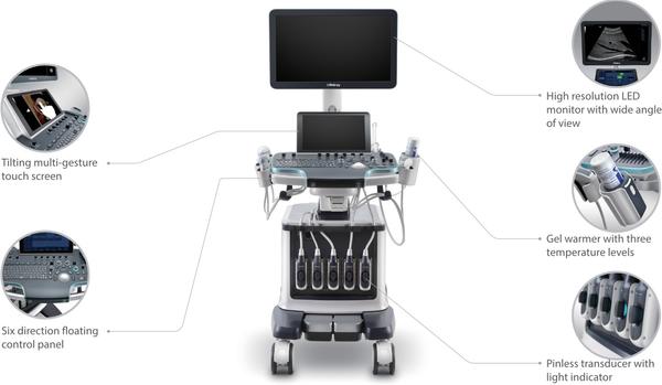
 Ergonomics The Resona 7 is designed around you. Gesture-based operation opens up a new trend in cart-based ultrasound with an agile, smart, and intuitive user experience beyond your expectations. Gel warmer’s three level temperature and swiveling angle adjustment of the control panel delivers great patient comfort and user convenience.

Send Message
product image
Hemoglobin Meter

Get the fastest hemoglobin and HCT test within 10 seconds, Buy Accurex URIT-12 Hemoglobin Meter Online. This Hemoglobin tester gives Accurate & Reliable Result. The Urit 12 hemoglobin machine is the best digital hemoglobin Meter with easy 3-step operation. The hb meter is is perfectly fit for the filtration of the numbers of blood donors, POCT, and hemoglobin testing in medical examination center. etc. It works on the prinicple of Optical reflectance. Sample: Capillary or venous whole blood (13~15 ul) & Strips: Accurex H12 Hemoglobin Test Strip URIT-12 is an easy-operation, fast-testing and portable hemoglobin testing Meter, which is perfectly fit for the filtration of the numbers of blood donors, POCT, and hemoglobin testing in medical examination center (MEC) etc. Compared to HiCN-HB method, the results are satisfactory. • Fast hemoglobin and HCT test within 10 seconds • Accurate & Reliable Result • Easy 3-step operation Parameter: Hemoglobin, HCT (Hematocrit) Principle: Optical reflectance Strip: H12 Hemoglobin Test Strip Speed: 10 seconds Sample: Capillary or venous whole blood (13~15 ul) Memory: 250 test results Measuring Range: 4.0g/dL 24.0g/dL(g/L optional) Quality Control: HQ-A Hemoglonbin control solution Calibration: Factory calibrated according to ICSH Power: DC 6V (Two CR2032 Batteries) Automatization: Self-checking, auto-judging and displaying malfunction Dimension: 102mm 50mm 19mm Accuracy: accuracy ≥ 96%,the correlation is very good in the comparison between URIT-12 and ICSH method. Precision: CV < 5%,testing samples including high, medium & low concentration, the CV of 10 repeated test results is 1.8% 2.2% 2.7% respectively. Hematology analyzer is not suitable for the fast and single hemoglobin testing out of the laboratory and the fast hemoglobin filtration for numbers of blood donors, therefore, it’s very necessary to find a kind of method for fast and accurate hemoglobin testing. When a drop of whole blood is applied to the test spot on the strip, blood immediately disperses within the membrane, contacting the reagent. The meter’s optical detectors automatically measure the change of reflectance in membrane. The intensity of reflectance is inversely proportional to the hemoglobin concentration. Other Info Hemoglobin testing is one of the important clinical hemanalysis items. It is useful for the diagnosis of all kinds of anemia. In clinical laboratory, many test items including hemoglobin can be gotten from hematology analyzer.

Send Message
product image
Rapid Test

Buy AccuTest Typhoid IgG/IgM Rapid Card Test (5 Tests) Online from Accurex. The Typhoid IgG/IgM Rapid Test is used for detection & differentiation of anti-Salmonella typhi (S. typhi) IgG and IgM in human whole blood / serum / plasma. This Typhoid test is used as a screening test and as an aid in the diagnosis of infection with S. typhi. Salmonella typhi bacteria causes typhoid Typhoid fever, typhoid symptoms symptoms are likely to develop gradually – appearing one to three weeks after exposure to the disease. There are various typhoid treatment & typhoid vaccine which can be opted after the detetction of Typhoid. Typhoid fever is caused by S. typhi, a Gram-negative bacterium. Worldwide an estimated 17 million cases and 600,000 associated deaths occur annually. Patients who are infected with HIV are at significantly increased risk of clinical infection with S. typhi. Evidence of H. pylori infection also presents an increased risk of acquiring typhoid fever. 1-5% of patients become chronic carrier harboring S. typhi in the gall bladder. The clinical diagnosis of typhoid fever depends on the isolation of S. typhi from blood, bone marrow or a specific anatomic lesion. In the facilities that can not afford to perform this complicated and time-consuming procedure, Filix-Widal test is used to facilitate the diagnosis. However, many limitations lead to difficulties in the interpretation of the Widal test. In contrast, the Typhoid IgG/IgM Rapid Test is a simple and rapid laboratory test. The test simultaneously detects and differentiates the IgG and the IgM antibodies to S. typhi specific antigen thus to aid in the determination of current or previous exposure to the S. typhi. PRINCIPLE The Typhoid IgG/IgM Rapid Test is a lateral flow immunochromatographic assay. The test cassette consists of: 1) a pink-purple colored conjugate pad containing recombinant S. typhi H antigen and O antigen conjugated with colloid gold (Typhoid conjugates) and rabbit IgG-gold conjugates, 2) a nitrocellulose membrane strip containing two test region (Test line M and G) and a control region (control line). The test line ‘M’ is pre-coated with monoclonal anti-human IgM for the detection of IgM anti-S. typhi, test line ‘G’ is pre-coated with monoclonal anti-human IgG for the detection of IgG anti-S. typhi , and the C band is pre-coated with goat anti rabbit IgG. When an adequate volume of test specimen is dispensed into the sample well of the cassette, the test specimen migrates by capillary action across the test cassette. Anti-S. typhi IgM if present in the patient specimen will bind to the Typhoid conjugates. The immunocomplex is then captured on the membrane by the pre-coated anti-human IgM antibody at test region ‘M’, forming a pink-purple line at ‘M’ region, indicating a S. typhi IgM positive test result. Anti-S. typhi IgG if present in the patient specimen will bind to the Typhoid conjugates. The immunocomplex is then captured by the pre-coated reagents on the membrane, forming a pink-purple line at test region ‘G’, indicating a S. typhi IgG positive test result. Absence of any pink-purple line at test region (M and G) suggests a negative result. The test contains an internal control (control line) which should exhibit a pink-purple line of the immunocomplex of goat anti rabbit IgG/rabbit IgG-gold conjugate regardless of the color development on any of the test region. Otherwise, the test result is invalid and the specimen must be retested with another device. PRECAUTION 1. This package insert must be read completely before performing the test. Failure to follow the insert gives inaccurate test results. 2. Do not open the sealed pouch, unless ready to conduct the assay. 3. Do not use expired devices. 4. Bring all reagents to room temperature (15°C-30°C) before use. 5. Do not use the components in any other type of test kit as a substitute for the components in this kit. 6. Do not use hemolized blood specimen for testing. Wear protective clothing and disposable gloves while handling the kit reagents and clinical specimens. Wash hands thoroughly after performing the test. 7. Users of this test should follow the US CDC Universal Precautions for prevention of transmission of HIV, HBV and other blood-borne pathogens. 8. Do not smoke, drink, or eat in areas where specimens or kit reagents are being handled. 9. Dispose of all specimens and materials used to perform the test as biohazardous waste. 10. Handle the Negative and Positive Control in the same manner as patient specimens. 11. The testing results should be read within 15 minutes after a specimen is applied to the sample well or sample pad of the device. Read result after 15 minutes may give erroneous results. 12. Do not perform the test in a room with strong air flow, ie. an electric fan or strong air-conditioning. Other Info STORAGE AND STABILITY Typhoid IgG/IgM test card should be stored at 4-40°C. The card may be stored at room temperature but not exceeding 40°C in the original sealed pouch. The shelf life or expiry of the card is printed on Pouch as well as carton label. The test kit should be kept away from direct sunlight, moisture and heat.

Send Message
product image
Rapid Test

Buy AccuTest H Pylori AB Rapid Card Test (50 Tests) Online from Accurex. The Helicobacter Pylori is a lateral flow chromatographic immunoassay for the qualitative detection of antibodies to H.pylori in human Whole Blood/Serum/Plasma. H Pylori test provides an aid in the diagnosis of infection with H.pylori. H.pylori is implicated in the etiology of a variety of gastrointestinal diseases, including duodenal and gastric ulcer, non-ulcer dyspepsia and active and chronic gastritis. h pylori symptoms are an ache or burning pain in your stomach (abdomen), Nausea, Loss of appetite, Frequent burping, Bloating & Unintentional weight loss NTENDED USE The H.pylori Antibody Rapid Test Card is a lateral flow chromatographic immunoassay for the qualitative detection of antibodies to H.pylori in human Whole Blood/Serum/Plasma. It provides an aid in the diagnosis of infection with H.pylori. INTRODUCTION H.pylori is implicated in the etiology of a variety of gastrointestinal diseases, including duodenal and gastric ulcer, non-ulcer dyspepsia and active and chronic gastritis. H.pylori infection is found in more than 90% of duodenal ulcer patients and in around 75% of all peptic ulcer sufferers. H.pylori infection is also more common in gastric cancer patients. The risk of gastric cancer has been estimated to be six-fold higher in H.pylori infected populations than in uninfected populations. H.pylori infections occur in human populations throughout the world. In developed countries, about 50% of the population may have H.pylori infection by the age of 60 years, while only 10-20% of adults in the third decade of life have it. Transmission is most probably by the fecal-oral or oral-oral route. Both invasive and non-invasive methods are used to diagnose H.pylori infection in patients with symptoms of gastrointestinal disease. Invasive methods include culture of gastric biopsy samples, histologic examination of stained biopsy specimens, or direct detection of the urease activity in the biopsy (CLO test). These methods need to obtain a biopsy sample by endoscopy.which is expensive,and usually results discomfort and risk to the patient.noninasive techniques include urea breath tests and serological methods. Urea breath test requires expensive laboratory equipment and moderate radiation exposure. Serologic tests are employed to detect antibodies as human immune response to H.pylori, for example the ELISA and the Western immunoblot. The H.pylori Antibody Rapid Test Card detect antibodies to H.pylori infection in human Whole Blood/Serum/Plasma. It is a noninvasive method and does not use radioactive isotopes. The test is easy to perform and requires no specialized equipment. Visual interpretation provides an accurate qualitative result. It is a useful on-site aid in the diagnosis of H.pylori infection. Diagnosis of H.pylori infection by antibody immunoassay can reduce the number of patients requiring endoscopy. PRINCIPLE The H.pylori Antibody Rapid Test Card is an immunoassay based on the principle of the double antigen-sandwich technique. During testing, a Whole Blood/Serum/Plasma specimen migrates upward by capillary action. The antibodies to H.pylori if present in the specimen will bind to the H.pylori conjugates. The immune complex is then captured on the membrane by the pre-coated H.pylori antigens, and a visible pink-purple line will show up in the test line region indicating a positive result. If antibodies to H.pylori are not present or are present below the detectable level, a pink-purple line will not form in the test line region indicating a negative result. To serve as a procedural control, a pink-purple line will always appear at the control line region, indicating that proper volume of specimen has been added and membrane wicking has occurred. Safety Information The H.pylori Antibody Rapid Test Card is limited to provide a qualitative detection. The intensity of the test line does not necessarily correlate to the concentration of the antibody in the blood. x The results obtained from this test are intended to be an aid in diagnosis only. Each physician must interpret the results in conjunction with the patient’s history, physical findings, and other diagnostic procedures. x A negative test result indicates that antibodies to H.pylori are either not present or at levels undetectable by the test. Direction for Use TEST PROCEDURE 1. Remove the test cassette from the sealed pouch and use it as soon as possible. 2. Place the test cassette on a clean and level surface. 3. For serum or plasma specimen: Hold the dropper vertically and transfer 3 drops of serum or plasma to the specimen well (S) of the test cassette, then start the timer. See illustration below. 4. For whole blood specimens: Hold the dropper vertically and transfer 1 drop of whole blood (approximately 30μl) to the specimen well(S) of the test cassette, then add 2 drops of buffer (approximately 70μl) and start the timer. 5. Read results at 15 minutes. Do not interpret the result after 20 minutes. Other Info STORAGE AND STABILITY x Store as packaged in the sealed pouch at 4-40℃. The kit is stable within the expiration date printed on the label. x Once open the pouch, the test should be used within one hour. Prolonged exposure to hot and humid environment will cause product deterioration.

Send Message

Still searching for
accurate diagnosis?