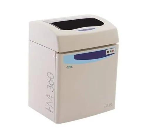
Jaipur
+918529833535

'60 tests'

Items tagged with '60 tests'

product image
Laboratory Usage

Using best-in-class technology, the Mindray CL-6000i is a chemiluminescence immunoassay analyzer with high throughput, high performance and high reliability. Features Throughput: up to 480 tests per hour Measurement principle: enhanced ALP-AMPPD method Reagent carousel: 36 reagent positions with non-stop refrigerating Sample handling: up to 300 samples can be loaded in one batch, sample loading and offloading continuously by sample racks, fast prioritizing STAT samples Sample volume: 5-110 μL Continuously loading of reagents, substrate, cuvettes, wash buffer and waste bags Zero daily maintenance Product Specification Model Name/Number CL-6000i Usage/Application Hospital Brand Mindray Sample Volume Range 5-110 microlitre Throughput up to 480 tests per hour Measurement Principle enhanced ALP-AMPPD method Reagent Carousel 36 reagent positions with non-stop refrigerating Sample Handling up to 300 samples can be loaded in one batch,sample loading and offloading continuously by sample r

Send Message
product image
Urinalysis Analyzer

High Performance "Full Automated System" Color Touch Panel for good usability 7 inch WVGA color touchscreen with high operability displays measurement results, instrument conditions and operating instructions. "Sample delivery system" enables micro volume samples to be measured As the sample is drawn through the nozzle and delivered correctly and quantitatively to each pad of the strip, the micro volume samples (minimum 1mL) can be measured easily. No cross contaminations are found on the reagent portions despite high sample volume. Sample Urine Test items Glu, Pro, Bil, Uro, Ket, pH, Nit, Bld, Leu, S.G., Alb, Cre(Alb/Cre ratio, Pro/Cre ratio can be calculated), turbidity and color tone Wavelength 5 LED wavelengths (430, 500, 565, 635, 760nm) Measurement principle Test strip :Dual-wavelength reflectance method (One-wavelength for Bld) Specific gravity :Reflection refractometryColor hue :Light-transmission measurementTurbidity :Light-scattering measurement method Test strip AUTION Sticks (10EA & 10PA) , Uriflet S 11UA Reaction time Approximately 60 seconds Throughput 225 samples per hour Strip storage Maximum 400 strips (200 strips x 2 feeder boxes) Required sample volume Normal, STAT measurement: Minimum 2 mL Small volume measurement: Minimum 1 mL Sample consumption Normal, STAT measurement: Maximum 0.90 mL Small volume measurement: Maximum 0.45 mL Sample container Sample tubes (length: 95 - 110 mm, diameter: ø14.0 - 15.8 mm) Maximum sample load 100 samples (Circulation) Data memory Normal, STAT, small volume measurement: 2500 tests Control measurement: 200 tests Check measurement: 50 tests Trouble list: 100 tests Display Large color liquid crystal display (800 × 480 dots) Built-in printer 58-mm width printer paper (24 digits) External output 1 port (Replaceable with the optional Ethernet terminal) Communication method RS-232C compliant (Switchable between one-way and two-way) Measurement environment Temperature: 10 - 30°C; Humidity: 30 - 60% RH (No condensation) Power requirements 100 - 240 V AC (Maximum power line fluctuation of ±10%), 50/60 Hz Power consumption 150VA (Maximum) Dimensions 530 (W) × 530 (D) × 550 (H) mm (including the sampler) Weight Main body: Approximately 36 kg Sampler: Approximately 4 kg

Send Message
product image
Rapid Test

Buy AccuTest Typhoid IgG/IgM Rapid Card Test (5 Tests) Online from Accurex. The Typhoid IgG/IgM Rapid Test is used for detection & differentiation of anti-Salmonella typhi (S. typhi) IgG and IgM in human whole blood / serum / plasma. This Typhoid test is used as a screening test and as an aid in the diagnosis of infection with S. typhi. Salmonella typhi bacteria causes typhoid Typhoid fever, typhoid symptoms symptoms are likely to develop gradually – appearing one to three weeks after exposure to the disease. There are various typhoid treatment & typhoid vaccine which can be opted after the detetction of Typhoid. Typhoid fever is caused by S. typhi, a Gram-negative bacterium. Worldwide an estimated 17 million cases and 600,000 associated deaths occur annually. Patients who are infected with HIV are at significantly increased risk of clinical infection with S. typhi. Evidence of H. pylori infection also presents an increased risk of acquiring typhoid fever. 1-5% of patients become chronic carrier harboring S. typhi in the gall bladder. The clinical diagnosis of typhoid fever depends on the isolation of S. typhi from blood, bone marrow or a specific anatomic lesion. In the facilities that can not afford to perform this complicated and time-consuming procedure, Filix-Widal test is used to facilitate the diagnosis. However, many limitations lead to difficulties in the interpretation of the Widal test. In contrast, the Typhoid IgG/IgM Rapid Test is a simple and rapid laboratory test. The test simultaneously detects and differentiates the IgG and the IgM antibodies to S. typhi specific antigen thus to aid in the determination of current or previous exposure to the S. typhi. PRINCIPLE The Typhoid IgG/IgM Rapid Test is a lateral flow immunochromatographic assay. The test cassette consists of: 1) a pink-purple colored conjugate pad containing recombinant S. typhi H antigen and O antigen conjugated with colloid gold (Typhoid conjugates) and rabbit IgG-gold conjugates, 2) a nitrocellulose membrane strip containing two test region (Test line M and G) and a control region (control line). The test line ‘M’ is pre-coated with monoclonal anti-human IgM for the detection of IgM anti-S. typhi, test line ‘G’ is pre-coated with monoclonal anti-human IgG for the detection of IgG anti-S. typhi , and the C band is pre-coated with goat anti rabbit IgG. When an adequate volume of test specimen is dispensed into the sample well of the cassette, the test specimen migrates by capillary action across the test cassette. Anti-S. typhi IgM if present in the patient specimen will bind to the Typhoid conjugates. The immunocomplex is then captured on the membrane by the pre-coated anti-human IgM antibody at test region ‘M’, forming a pink-purple line at ‘M’ region, indicating a S. typhi IgM positive test result. Anti-S. typhi IgG if present in the patient specimen will bind to the Typhoid conjugates. The immunocomplex is then captured by the pre-coated reagents on the membrane, forming a pink-purple line at test region ‘G’, indicating a S. typhi IgG positive test result. Absence of any pink-purple line at test region (M and G) suggests a negative result. The test contains an internal control (control line) which should exhibit a pink-purple line of the immunocomplex of goat anti rabbit IgG/rabbit IgG-gold conjugate regardless of the color development on any of the test region. Otherwise, the test result is invalid and the specimen must be retested with another device. PRECAUTION 1. This package insert must be read completely before performing the test. Failure to follow the insert gives inaccurate test results. 2. Do not open the sealed pouch, unless ready to conduct the assay. 3. Do not use expired devices. 4. Bring all reagents to room temperature (15°C-30°C) before use. 5. Do not use the components in any other type of test kit as a substitute for the components in this kit. 6. Do not use hemolized blood specimen for testing. Wear protective clothing and disposable gloves while handling the kit reagents and clinical specimens. Wash hands thoroughly after performing the test. 7. Users of this test should follow the US CDC Universal Precautions for prevention of transmission of HIV, HBV and other blood-borne pathogens. 8. Do not smoke, drink, or eat in areas where specimens or kit reagents are being handled. 9. Dispose of all specimens and materials used to perform the test as biohazardous waste. 10. Handle the Negative and Positive Control in the same manner as patient specimens. 11. The testing results should be read within 15 minutes after a specimen is applied to the sample well or sample pad of the device. Read result after 15 minutes may give erroneous results. 12. Do not perform the test in a room with strong air flow, ie. an electric fan or strong air-conditioning. Other Info STORAGE AND STABILITY Typhoid IgG/IgM test card should be stored at 4-40°C. The card may be stored at room temperature but not exceeding 40°C in the original sealed pouch. The shelf life or expiry of the card is printed on Pouch as well as carton label. The test kit should be kept away from direct sunlight, moisture and heat.

Send Message
product image
Rapid Test

Buy AccuTest HBsAg Rapid Card Test (50 Tests) Online from Accurex. HBsAg means Hepatitis B surface antigen which is a protein on the surface of hepatitis B virus. It can be detected in high levels in serum during acute or chronic hepatitis B virus infection. HBsAg positive indicates that the person is infectious. The hbsag test kit is a one-step immunochromatographic assay based on the antigen capture, or Sandwich principle. HBsAg positive treatment may include Antiviral medications. AccuTest HBsAg Card Test Hepatitis BHepatitis B is a viral infection that attacks the liver and can cause both acute and chronic disease. An estimated 240 million people are chronically infected with hepatitis B (defined as hepatitis B surface antigen positive for at least 6 months). Laboratory diagnosis of hepatitis B infection focuses on the detection of the hepatitis B surface antigen HBsAg. Hepatitis B surface antigen (HBsAg) consists of lipid carbohydrate and protein elements; the protein moiety provides a marker for identification of chronic, infectious BBV infections. Hepatitis B is transmitted sexually or intravenously and has an incubation period of six months. If not diagnosed properly and in time, it can develop into acute or chronic infection, liver cirrhosis and fulminant hepatitis. This test is very useful for screening blood donors, to find out whether they are HBsAg positive before collection of blood. Principle: AccuTest HBsAg is a qualitative test based on immunochromatography sandwich principle. The test card includes a combination of monoclonal anti-body gold conjugate (colloidal gold) and polyclonal solid phase antibodies which selectively binds Hepatitis B surface antigen with high degree of sensitivity. The HBsAg test is a one-step immunochromatographic assay based on the antigen capture, or Sandwich principle. The method uses monoclonal antibodies conjugated to colloidal gold and polyclonal antibodies immobilized on a nitrocellulose strip in a thin line. The test sample is introduced into well and flows laterally through an absorbent pad where it mixes with the signal reagent. If sample contains HBsAg, the colloidal gold-antibody (mouse) conjugate binds to the antigen, forming an antigen-antibody-colloidal gold complex. The complexes then migrate through the nitrocellulose strip by capillary action, which are stopped by an immobilized antibody zone forming a purple band. The formation of the first purple band (T zone) is indicative of hepatitis positive. To serve as a procedural control, an additional line of antibody antimouse IgG has been immobilized on the card. If the test is performed correctly, this will result in the formation of purple and upon contact with the conjugate as a control line. Safety Information AccuTest HBsAg test kit is designed lor detection of Hepalitis B Surface Antigenin the specimen. As with all diagnostic tests, il is recommended to interpret all the results in line wilh clinical manifestations, other lest resulls & clinicians’s view colleclively. If the lest device is showing stagnancy in flow on chromalography, it is most likely due lo problem in the sample. In this case, relest with a fresh sample or diluted sample. Direction for Use Test Procedure: 1. Allow the test, specimen and or control to room temperature prior to the testing 2. Remove one test card from the pouch and place it on a clean flat surface 3. Using the dropper provided one drop of serum/plasma or W. blood sample (25 ul), wait for few seconds till the Whole blood absorbed by sample pad (approx. 30 second), 4. Add 2-3 drops of buffer (60 ul) into the sample well. (If needed add one more drop of assay buffer). 5. Let reaction proceed until the appearance of positive line and control line or up to 20 minutes. 6. Read results after 20 minutes, strong positive reaction may visible within 5 minutes. 7. If negative or questionable results are obtained, the HBV infection is suspected, the test should be repeated ona fresh serum specimen. Storage: AccuTest HBsAg should be stored at 4-30C. However, the card may be stored at room temperature not exceeding 30C in the original sealed pouch.

Send Message
product image
Rapid Test

Buy AccuTest H Pylori AB Rapid Card Test (50 Tests) Online from Accurex. The Helicobacter Pylori is a lateral flow chromatographic immunoassay for the qualitative detection of antibodies to H.pylori in human Whole Blood/Serum/Plasma. H Pylori test provides an aid in the diagnosis of infection with H.pylori. H.pylori is implicated in the etiology of a variety of gastrointestinal diseases, including duodenal and gastric ulcer, non-ulcer dyspepsia and active and chronic gastritis. h pylori symptoms are an ache or burning pain in your stomach (abdomen), Nausea, Loss of appetite, Frequent burping, Bloating & Unintentional weight loss NTENDED USE The H.pylori Antibody Rapid Test Card is a lateral flow chromatographic immunoassay for the qualitative detection of antibodies to H.pylori in human Whole Blood/Serum/Plasma. It provides an aid in the diagnosis of infection with H.pylori. INTRODUCTION H.pylori is implicated in the etiology of a variety of gastrointestinal diseases, including duodenal and gastric ulcer, non-ulcer dyspepsia and active and chronic gastritis. H.pylori infection is found in more than 90% of duodenal ulcer patients and in around 75% of all peptic ulcer sufferers. H.pylori infection is also more common in gastric cancer patients. The risk of gastric cancer has been estimated to be six-fold higher in H.pylori infected populations than in uninfected populations. H.pylori infections occur in human populations throughout the world. In developed countries, about 50% of the population may have H.pylori infection by the age of 60 years, while only 10-20% of adults in the third decade of life have it. Transmission is most probably by the fecal-oral or oral-oral route. Both invasive and non-invasive methods are used to diagnose H.pylori infection in patients with symptoms of gastrointestinal disease. Invasive methods include culture of gastric biopsy samples, histologic examination of stained biopsy specimens, or direct detection of the urease activity in the biopsy (CLO test). These methods need to obtain a biopsy sample by endoscopy.which is expensive,and usually results discomfort and risk to the patient.noninasive techniques include urea breath tests and serological methods. Urea breath test requires expensive laboratory equipment and moderate radiation exposure. Serologic tests are employed to detect antibodies as human immune response to H.pylori, for example the ELISA and the Western immunoblot. The H.pylori Antibody Rapid Test Card detect antibodies to H.pylori infection in human Whole Blood/Serum/Plasma. It is a noninvasive method and does not use radioactive isotopes. The test is easy to perform and requires no specialized equipment. Visual interpretation provides an accurate qualitative result. It is a useful on-site aid in the diagnosis of H.pylori infection. Diagnosis of H.pylori infection by antibody immunoassay can reduce the number of patients requiring endoscopy. PRINCIPLE The H.pylori Antibody Rapid Test Card is an immunoassay based on the principle of the double antigen-sandwich technique. During testing, a Whole Blood/Serum/Plasma specimen migrates upward by capillary action. The antibodies to H.pylori if present in the specimen will bind to the H.pylori conjugates. The immune complex is then captured on the membrane by the pre-coated H.pylori antigens, and a visible pink-purple line will show up in the test line region indicating a positive result. If antibodies to H.pylori are not present or are present below the detectable level, a pink-purple line will not form in the test line region indicating a negative result. To serve as a procedural control, a pink-purple line will always appear at the control line region, indicating that proper volume of specimen has been added and membrane wicking has occurred. Safety Information The H.pylori Antibody Rapid Test Card is limited to provide a qualitative detection. The intensity of the test line does not necessarily correlate to the concentration of the antibody in the blood. x The results obtained from this test are intended to be an aid in diagnosis only. Each physician must interpret the results in conjunction with the patient’s history, physical findings, and other diagnostic procedures. x A negative test result indicates that antibodies to H.pylori are either not present or at levels undetectable by the test. Direction for Use TEST PROCEDURE 1. Remove the test cassette from the sealed pouch and use it as soon as possible. 2. Place the test cassette on a clean and level surface. 3. For serum or plasma specimen: Hold the dropper vertically and transfer 3 drops of serum or plasma to the specimen well (S) of the test cassette, then start the timer. See illustration below. 4. For whole blood specimens: Hold the dropper vertically and transfer 1 drop of whole blood (approximately 30μl) to the specimen well(S) of the test cassette, then add 2 drops of buffer (approximately 70μl) and start the timer. 5. Read results at 15 minutes. Do not interpret the result after 20 minutes. Other Info STORAGE AND STABILITY x Store as packaged in the sealed pouch at 4-40℃. The kit is stable within the expiration date printed on the label. x Once open the pouch, the test should be used within one hour. Prolonged exposure to hot and humid environment will cause product deterioration.

Send Message
product image
Hematology

Buy the Best 5 Parts Hematology Analyzer – ACCULAB CBC 560+. This Hematology Analyser Machine is an advanced and automatic hematology analyzer, comprising 5 parts, 29 parameters, and 3 histograms assay, offers throughput of 60 tests/hour. Features: Tri-angle laser scatter, Flow cytometry method, Impedance method for RBC and PLT counting and Cyanide-free method for HGB test along with 3D holographic scattergram to display the accurate 5 part differentiation of WBC. Equipped with a 14 inch touch screen and large storage capacity for 100,000 results, this hematology analyzer has interface of 4 USB ports, 1 LAN port Bi-direction LIS, support HL 7 protocol, Internal RFID reader. With CBC mode, CBC+DIFF mode, Venous whole blood, Capillary whole blood and Prediluted test mode, this hematology analyzer is an ideal unit for white blood cell counts, complete blood Measuring principle Tri-angle laser scatter, Flow cytometry method, Impedance method for RBC and PLT counting, Cyanide-free method for HGB test Sample volumes CBC+DIFF mode: ≤ 20 μl CBC mode: ≤ 10 μl Throughput 60 tests/hour Assay Items 29 parameters, 3 histograms Display 14 inch touch screen Storage 100,000 sample results, including histograms, scattergrams and patient information Parameter WBC, RBC, HGB, HCT, MCV, MCH, MCHC, RDW-SD, RDW-CV, PLT, MPV, PCT,PDW, P-LCR, P-LCC, NEU%, LYM%, MON%, EOS%,BAS%,NEU#, LYM#, MON#, EOS#, BAS# 4 Research parameter: ALY%, ALY#, LIC%, LIC# Performance Item Linearity range Carry Over CV WBC 0-300 × 109/L ≤ 0.5% ≤ 2.0 % RBC 0-8.00 ×10¹²/L ≤ 0.5% ≤ 1.5 % HGB 0-250 g/L ≤ 0.5% ≤ 1.5 % PLT 0-3000 × 109/L ≤ 1.0% ≤ 4.0 % Interface 4 USB ports, 1 LAN port Bi-direction LIS, support HL 7 protocol, Internal RFID reader Power Consumption 400 VA Power Supply AC 220V±10%, 50/60Hz, 110V±10%, 60Hz Packaging dimension (W×D×H) 670 × 590 × 790 mm Gross Weight 53 kg Safety Information All the samples, controls, calibrators, reagents, waste and areas contacted with them are potentially biohazardous. Wear proper personal protective equipment (e.g. gloves, lab coat, etc.) and follow safe laboratory procedures when handling them in the laboratory.

Send Message
product image
Clinical Chemistry

The HumaStar 600 is designed for being on duty 24/7 to run your daily photometric and immunoturbidimetric routine as well as optional direct ISE measurement. It incorporates a state-of-the-art technology with sophisticated but versatile and user-friendly software. 900 to 2250 tests / day Water consumption < 5l/h Up to 600 chemistry tests per hour (w/o ISE) Up to 780 tests per hour with optional ISE module 48 cooled reagents on board Reagent cooling Continous programming and sample loading Barcode reader for samples and reagents

Send Message
product image
Hemostasis Analyzer

ECL 760 ECL 760 is an advanced Fully automated coagulation analyzer with seven detection channels providing operators with a compact easy-to-use system. Dedicated for mid-volume throughput labs, the ECL 760 been designed to optimize your productivity. ECL 760 is a blend of modernity and simplicity. Erba reagents oers a wide menu of test parameters for automated coagulation analyzers. KEY FEATURES ECL 760 uses the proven system of percentage intensity of light scatter at 640 nm LED. This optical system ensures early light stability, longer duration usage time and greater lamp life. It minimizes the interference of hemolysis, ictericity and lipemia, in samples at each assay, The system monitors the entire clotting process from reagent addition to complete clot formation with the generation of Clot curve. Clotting Tests  All clotting tests, such as Prothrombin Time, Activated Partial Thromboplastin Time, Fibrinogen, Thrombin time, Factors (II / V / Vll / X / Vlll / IX\ Xl / Xll), Lupus and Protein S are performed. Chromogenic Tests  Chromogenic tests use the colorimetric principle of measuring absorbance of light (405nm LED) by the test in a cuvette. Protein C and Antithrombin Illare some of the tests done by this method. Immunological Assay  The change in light intensity caused by the antigen antibody reaction is detected at 575 nm LED, as the change in the transmitted light.  D Dimer is the most common test performed

Send Message
product image
Clinical Chemistry

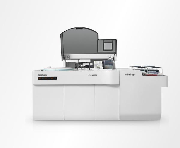
EM 360 is a Fully Automated Clinical Chemistry Analyser which is time tested and proven technology. It is designed to provide the most demanding productivity, speed and operational ease. EM 360 with a throughput of 360 tests/hour offers flexibility in operation and rugged performance to win against high workload, tight deadlines, multiple tasks and complexity of testing. It yields high quality results for virtually any biochemistry test to deliver quick, efficient and consistent performance. Product Features: Automated, Discrete, Patient prioritized, Floor model Based on proven technology, Diffraction Grating Separate probes for Reagent R1 & R2 and dedicated probe for sample Low reading volume of 200 µl only Onboard cooling for reagents to enhance the reagent stability Barcode for reagents & samples (Optional) Wide test menu Product Applications: End Point Reactions: Glucose, Cholesterol, Triglycerides, Albumin, Total Protein, Uric Acid, Calcium, Magnesium, Bilirubin Total, Bilirubin Direct Kinetic Reactions: SGOT, SGPT, GGT, LDH, CK, CK MB, ALP, ACP, Amylase, Lipase Two Point Rate Reactions: Urea, Creatinine Immunoturbidimetric Chemistries: RA, CRP, ASO, Direct HDL, Direct LDL, Apo A, ApoB, Lp (a) Special Chemistries: HbA1c, Iron, Ferritin, Phosphorus, Microprotein, Ammonia, Bicarbonates System Type: Discrete, automated, random access, patient prioritized clinical chemistry analyzer Throughput: 360 tests / hour photometric and 600 tests / hour with ISE* (*optional ISE with Na+, K+, Cl-, Li+) Analytical Methods: 1-Point, 2-Point, Rate-A, Rate-B, Direct Potentiometry (optional) Barcode Reader: For reagents and samples (optional) Reaction Tray: 60 hard glass cuvettes Reaction Mixing: Stirrer with variable speed Minimum reaction Volume: 150 μl with maximum 200 μl reading volume On-board Laundry: 5 stage cleaning, 2 stage drying Photometer: Multi-wavelength diffraction grating with 12 wavelengths (340, 376, 415, 450, 480, 505, 546, 570, 600, 660, 700, 750 nm) OD Range: 0.0 - 3.0 Light Source: Halogen lamp Detector: Silicon photo-diode Water Consumption: < 10 litres Programmable Parameters: Default system pack parameters + upto 99 user defined parameters, Unlimited profile and unlimited calculation items Quality Control: QC plot data with QC rules. Provision for lab mean. Twin plot Calibration: K-Factor, Linear (1, 2 point & multipoint), 4P and 5P Logit-log, cubic spline, exponential, polynomial, On-board serial dilution for calibrator Power Requirement: AC 220 V ± 10 %, 50 Hz or AC 110 V ± 10 %, 60 Hz Power Consumption: 800 VA Dimension (mm): Approx. 675 (W) x 840 (D) x 1120 (H) Weight: Approx. 150 kgs. SAMPLE HANDLING Sample: Serum, Plasma, Urine, CSF, Whole blood, other Sample Unit: 82 positions for samples, blank , controls, calibrators, STAT sample Sample Pipetting: 2 - 70 µl (adjustable in 0.1 µl step) for Biochemistry, 70 µl fixed for ISE Capacitance probe with liquid level sensing & vertical obstruction detection, serum indices Auto Rerun: Repeat with same, increased or decreased volume ( upto 1:150) Sample Tubes /Cups: Primary tubes of 5 ml, 7 ml, 10 ml and sample cups REAGENT HANDLING Reagent Tray: 50 positions for reagents with onboard cooling Reagent Pipetting: R1: 50 - 300 µl (adjustable in 1 µl step) R2: 10 - 200 µl (adjustable in 1 µl step) Capacitance probe with level sensing & vertical obstruction detection SYSTEM INTERFACE: Analyzer-PC: USB PC - Host Computer: Bidirectional TCP / IP & RS - 232 PC - Printer: USB Operating System: Windows Based Database: Unlimited Results

Send Message

Still searching for
60 tests?